Search This Blog

Sunday, December 17, 2006

Somatostatin Receptor Imaging and Small Cell Lung Ca

Tc99m Depreotide Scan (NeoTect): Small cell lung cancer in the left lower lobe with possible metastatic left axillary lymph node. There is some more subtle mediastinal activity as well.
Somatostatin is a 14-amino-acid peptide hormone found on many cells of neuroendocrine origin. It acts as a neurotransmitter in the central nervous system. When it binds to cells, it inhibits the release of growth hormone, insulin, glucagon, and gastrin. Somatostatin receptors have been found on the surface of many human tumor cells including APUD cells (amine precursor uptake and decarboxylation) such as pituitary tumors, pancreatic endocrine tumors, carcinoids, paragangliomas, small cell lung cancers, medullary thyroid carcinomas and pheochromocytomas. Small cell lung cancer (SCLC) tumors have neuroendocrine features. In vitro and in vivo studies have demonstrated that 50%-75% of SCLC tumors express receptors for somatostatin. Non-APUD tumor cells have somatostatin receptors, including activated lymphocytes, astrocytomas, and some breast carcinoma. Studies have shown that somatostatin analogs may inhibit growth of many of these tumors in vivo in animal studies. Tumors such as non small cell lung cancer that do not express somatostatin receptors are nonetheless imaged. One possible explanation of this finding is that the uptake of tracer is not by the receptor-negative tumor but by the surrounding tissues such as somatostatin receptor-positive white blood cells or neuroendocrine cells nearby the primary tumors. In-111-DTPA-D-Phe1]-octreotide was found to have a high affinity for somatostatin receptors . The compound, also called OctreoScan, is easily labeled with In-111. Since this radiotracer is mainly eliminated via the kidneys, intra-abdominal evaluation of somatostatin-receptor positive tumors could be performed. Tc99m Depreotide (NeoTect) is a somatostatin peptide analog labled with Technetium. FDG PET has decreased the use of somatostatin receptor scintigraphy for lung cancer.


Tuesday, December 12, 2006

Neurofibrosarcoma in NF1 Patient. PET / MR Fusion

Click on Image for Larger View

Young adult female with known NF1 and multiple peripheral neuromas. The patient had a prior neurofibrosarcoma in the left sacral plexus resected 4 years ago. She presented to another hospital for pain and on a routine MRI of the lumbar spine a suspicicious area was noted. PET / CT was done at our facility and I fused it to the T2 axial Lsp MRI above. The patient has a recurrent neurofibrosarcoma in the left sacral plexus.

Friday, November 24, 2006

Type A Aortic Disection with Pericardial Hematoma

CT chest: Type A Aortic disection with rupture into pericardial space. Note the intimal flap in the ascending aorta and the intermediate density blood in the pericardial space.
Click image for full size

Pathophysiology
tear in intimal lining
ascending (65%)
arch (10%)
descending (20%)
Why?
collagen elastin abnormality
cystic medial necrosis
Intramural hematoma
No intimal tear
medial hemorrhage
Who?
HTN
vasculitis
50-70 yo males >females
Diagnosis
CT scan
sensitivity 96-100%
specificity 96-100%
Ptifalls- motion thru aortic root (can ecg gate)
renal insufficiency
contrast allergy
MRI
sens= 98%
spec= 98%
Pitfalls long study not for unstable patient
no iodine
less motion artifact if ecg gated
can quantitiate aortic insufficiency
Complications
Death: 25% first 24 hrs
50% first week
Aortic insufficiency
pericardial hematoma and tamponade
MI
branch artery occlusion
rupture leak
Famous deaths
King George II
John Ritter


Wednesday, November 15, 2006

Bone Scintigraphy Identifies Potential Responders to Plantar Fasciitis Treatment

Scan classification showing blood-pool phase at top and delayed phase at bottom: focal hyperemia in plantar fascia and marked delayed uptake in inferior calcaneum (A), hyperemia in proximal third and mild delayed uptake (B), and diffuse hyperemia and moderate delayed uptake (C). Arrowheads indicate hyperemia; arrows indicate delayed uptake. Copyright © 2006 by the Society of Nuclear Medicine.

Scintigraphy pegs potential responders to plantar fasciitis treatment
10/25/2006By: Plantar fasciitis is the most common cause of heel pain but the differential diagnosis can include Achilles tendinitis, retrocalcaneal bursitis, and calcaneum fracture. Bone scintigraphy is often used to identify the source of heel pain, and the modality may play a predictive role in treatment as well, according to a study in the Journal of Nuclear Medicine.

"Injections of corticosteroids or local anesthetics remain a convenient form of therapy," wrote Dr. Clayton Frater and colleagues. "Bone scintigraphy has also been shown to be an accurate guide in selecting injection sites.... We present scintigraphic criteria that can help identify potential responders (to therapy)..." (JNM, October 2006, Vol. 47:10, pp. 1577-1580).
Frater is from the University of Sydney in Australia. Other co-authors are from the University of New South Wales and Concord Hospital, both in Sydney, as well as King's College in London.
For this study, scintigraphy scans from 24 patients diagnosed with plantar fasciitis, and treated with local injection of steroids or anesthetics, were included. In addition, scans from 10 patients with other conditions were thrown into the mix to test the specificity of scintigraphy.

Imaging was done with a high-resolution collimator affixed to a gamma camera (Millennium or 400AC, GE Healthcare, Chalfont St. Giles, U.K.) after a 900- to 1,000-MBq injection of Tc-99m methylene diphosphonate. Blood-pool images of the plantar aspect of both feet were obtained, as were delayed images of the anterior, posterior, lateral/medial, and plantar aspects of the feet.
Patient responses were followed up at four to five weeks. Abnormalities of the blood-pool phase were characterized as focal calcaneal hyperemia, extension into the proximal third of the plantar fascia, diffuse involvement of the plantar fascia, or no evidence of hyperemia within the plantar fascia. Delayed images were classified based on the degree of calcaneal uptake.
In total, 32 injections were performed in 24 patients with complete or near relief from pain reported in 20 feet. Of the 20 that responded to treatment, 14 feet showed focal hyperemia and six showed minimal extension, the authors stated, adding that they found a significant difference between patients with focal hyperemia and diffuse blood-pool abnormalities (p = 0.004).
On the delayed images of the 20 responders, eight feet demonstrated mild inferior calcaneal uptake, six showed moderate uptake, and six had severe uptake.

"The blood-pool studies had good reproducibility, with a k-value of 0.64 (95% confidence interval [CI]). Agreement on reporting the delayed images was also good, with a k-score of 0.66 (95% CI)," the authors wrote. Interobserver agreement was fair with a k-value of 0.55, they reported.
"Focal calcaneal hyperemia is clearly associated with a high success rate for injection, with extension of hyperemia into the proximal soft tissues being associated with a response in only 50% and diffuse hyperemia with no response," the authors concluded. Using imaging criteria, clinicians can rule out patients who would not benefit from injection therapy, they added.

I thought this might be of interest to Podiatry Residents who rotate through.

Source: Shalmali Pal. AuntMinnie.com staff writerOctober 25, 2006

Monday, November 13, 2006

There is a very excellent podcast related to Nuclear Medicine and Molecular Medicine that can be found at http://nucmedpodcast.blogspot.com/ as well as at Itunes. As stated on the home page this podcast is "intended to be a community podcast with you the nuclear medicine professionals and users as the driving force. Please email suggestions, audio comments, and volunteers for podcast interviews. nucmedpodcast@gmail.com" I have been a regular listener since episode 1. (Nov 05: congrats on one year anniversary) What I like best about the podcast is that it gives an international view of whats going on in NUCS which is nice for someone from North America to get exposure to.

Saturday, November 11, 2006

Lung CA II PET / CT


Click image for full size
This is just a quick update to show a VR PET /CT of a left hilar lung cancer. This is a PET image fused to an average intensity projection image. I think it demonstrates nicely the relationship of the tumor uptake to the airway and the left upper lobe post obstructive pneumonia

In an earlier post I mentioned Amide as a nice freeware for image fusion. I should give equal time to MIM. This CLEVELAND based company founded by Dennis Nelson PhD makes terrific visualization software for nuclear imaging and medical imaging in general. It is actually what we use most for daily PET/CT reading. The reason is that it is very intuitive and does a great job streamlining PET /CT work flow. They are also doing a great job integrating their software with PACS systems.


Saturday, November 4, 2006

VR PET/CT Tongue Base Cancer

Click image for full size

Volume rendered PET/CT. There is a mid line squamous cell Ca of the tongue base. There is no adenopathy. We are seeing an increase in the number of patients with head and neck Ca being referred for PET/CT. As stated in an earlier post PET/CT's impact on staging and initial therapy decisions is significant.

Tuesday, October 31, 2006

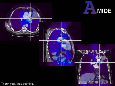
Image fusion, lung cancer, AMIDE

Click image for full size
In the old days before PET/CT, we did manly image fusion. We got separate dicom data from modalities thousands of miles apart and we tortured each voxel until they fit together as the fused image. It was fun. Now everything is done for you with PET/CT and excellent software like MIM. But every once and a while there is a rogue study. You get a stand alone PET scan from some outside site and a CT from somewhere else. What do you do?

Amide courtesy of Andy Loening at Sourceforge (amide.sourceforge.net) is a great open source software package for image fusion. It is available for Win32, Linux and MAC OSX (with some tweaking with x11 and fink). It is intuitive and robust. If you read PET from a standalone scanner or want to fuse PET/CT data to another (contrast enhanced?) CT or MRI this is an excellent option. Above is an example of a study from a standalone PET fused to a contrast enhanced CT from a different institution. There is a mass in the left hilum obstructing an upper lobe bronchus and causing post obstructive pneumonia. Fusion was performed with Amide.

This has me thinking that we should be performing fusion SPECT more often. We don't have a hybrid SPECT /CT camera and I doubt we will for some time, but Amide is an easy tool to use and won't add that much time for complex cases.

Monday, October 30, 2006

PET / CT Recurrent Laryngeal Carcinoma

Click image for full size

PET / CT has a greater sensitivity and specificity for detection of regional metastatic adenopathy for head and neck tumors than contrast enhanced CT. More importantly PET changes the course of management in patients with head and neck Ca in 33% of the cases where it is performed. It is also more sensitive and specific than CT for detection of local recurrence. There may be a role for PET in evaluating response to therapy for head and neck tumors but this is still developing. Remember many areas of normal FDG uptake are found in the head and neck including: Palatine, faucil and retropharyngeal tonsilar tissue, parotid, sub lingual and submandibular glands, strap muscles and the vocal cords.

Saturday, October 28, 2006

Follicular Cell Lymphoma of the Duodenum

Click on image for full size
This is a volume rendered PET/CT. Follicular cell extranodal lymphoma is rare in the duodenum where MALT would be more common. Follicular cell NHL is the most common indolent lymphoma and patients may go for years with no need of treatment. These tumors bear a chromasomal translocation which activates the bcl 2 gene. This protein blocks apoptosis aiding in cellular proliferation. Remember to be a cancer you not only have to proliferate, you have to circumvent cell death.

Monday, October 23, 2006

UCSF Womens Imaging Conference-Post Menopausal Bleeding and US


This slide from a lecture by Dr Ruth Goldstein is an elloquent summary of her talk on ultrasound and post menopausal bleeding. We don't know what endometrial thickness threshold to use in post menopausal women who are not bleeding but >99% will be normal with a thickness less than 8mm.

UCSF Womens Imaging Conference

Just returned from the UCSF Womens Imaging conference. Overall I would rate the meeting an 8/10. I will post one key concept from each of the 5 days of the conference over the next several entries.

Thursday, October 12, 2006

Klippel Trenaunay Syndrome

(Click image for full size)
Clinical triad of port wine nevus (hemangioma), lower extremity swelling and varicose veins. A congenital malformation of vascular and lymphatic structures usually in the lower extremities. Pateints develope lower extremity enlargement. May develope large lymphangiomas and hemangiomas.

Image A and D: Coronal and axial T2 fat supressed MRI of lower extremities in a 14m old male infant. Note the large perineal lymphatic mass extending upinto the pelvis. Also near the knees there are other vascular / lymphatic malformations.

Image B: Image fusion of contrast MRA with conventional T2 MRI in volume rendering. You can now see the full exrtent of the vascular / lymphatic malformations in 3D with ther relationship to the arteries. Note the limited visualization of lower extremity arteries in the left calf. Patients with KT syndrome can have arterial abnormalities including atresias in the lower extremities.

Image D: Fusion of MR venogram with T2 coronal images in volume rendering. Tee entire deep venous system has been replaced by extensive collaterals. The large lateral venous channels in the thighs are called Trenaunay veins.


Hangmans fracture 3D VR CT


Classic imaging findings for a Hangmans fracture of C2. The fractures plane extends from the posterior body of C2 into the posterior arch, in ths case through the pedicles. The fracture is unstable but often there is no initial cord injury since the fracture actually widens the canal. This fracture occurs with forced flexion, like that which occurs when the knot of a hangmans noose forces the chin up (hence the name).