 Click image for full size
Tuesday, October 31, 2006
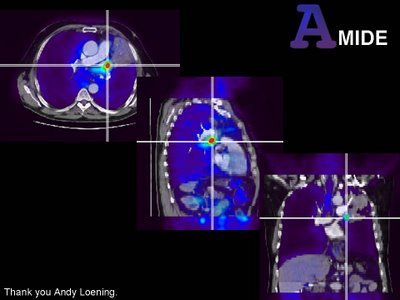
Image fusion, lung cancer, AMIDE
 Click image for full size
Monday, October 30, 2006
PET / CT Recurrent Laryngeal Carcinoma
 Click image for full size
PET / CT has a greater sensitivity and specificity for detection of regional metastatic adenopathy for head and neck tumors than contrast enhanced CT. More importantly PET changes the course of management in patients with head and neck Ca in 33% of the cases where it is performed. It is also more sensitive and specific than CT for detection of local recurrence. There may be a role for PET in evaluating response to therapy for head and neck tumors but this is still developing. Remember many areas of normal FDG uptake are found in the head and neck including: Palatine, faucil and retropharyngeal tonsilar tissue, parotid, sub lingual and submandibular glands, strap muscles and the vocal cords.
Saturday, October 28, 2006
Follicular Cell Lymphoma of the Duodenum
Monday, October 23, 2006
UCSF Womens Imaging Conference-Post Menopausal Bleeding and US
UCSF Womens Imaging Conference
Thursday, October 12, 2006
Klippel Trenaunay Syndrome
 (Click image for full size)
Clinical triad of port wine nevus (hemangioma), lower extremity swelling and varicose veins. A congenital malformation of vascular and lymphatic structures usually in the lower extremities. Pateints develope lower extremity enlargement. May develope large lymphangiomas and hemangiomas.
Image A and D: Coronal and axial T2 fat supressed MRI of lower extremities in a 14m old male infant. Note the large perineal lymphatic mass extending upinto the pelvis. Also near the knees there are other vascular / lymphatic malformations.
Image B: Image fusion of contrast MRA with conventional T2 MRI in volume rendering. You can now see the full exrtent of the vascular / lymphatic malformations in 3D with ther relationship to the arteries. Note the limited visualization of lower extremity arteries in the left calf. Patients with KT syndrome can have arterial abnormalities including atresias in the lower extremities.
Image D: Fusion of MR venogram with T2 coronal images in volume rendering. Tee entire deep venous system has been replaced by extensive collaterals. The large lateral venous channels in the thighs are called Trenaunay veins.
Hangmans fracture 3D VR CT

Classic imaging findings for a Hangmans fracture of C2. The fractures plane extends from the posterior body of C2 into the posterior arch, in ths case through the pedicles. The fracture is unstable but often there is no initial cord injury since the fracture actually widens the canal. This fracture occurs with forced flexion, like that which occurs when the knot of a hangmans noose forces the chin up (hence the name).


