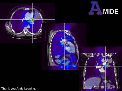
 Click image for full size
In the old days before PET/CT, we did manly image fusion. We got separate dicom data from modalities thousands of miles apart and we tortured each voxel until they fit together as the fused image. It was fun. Now everything is done for you with PET/CT and excellent software like MIM. But every once and a while there is a rogue study. You get a stand alone PET scan from some outside site and a CT from somewhere else. What do you do?
Amide courtesy of Andy Loening at Sourceforge (amide.sourceforge.net) is a great open source software package for image fusion. It is available for Win32, Linux and MAC OSX (with some tweaking with x11 and fink). It is intuitive and robust. If you read PET from a standalone scanner or want to fuse PET/CT data to another (contrast enhanced?) CT or MRI this is an excellent option. Above is an example of a study from a standalone PET fused to a contrast enhanced CT from a different institution. There is a mass in the left hilum obstructing an upper lobe bronchus and causing post obstructive pneumonia. Fusion was performed with Amide.
This has me thinking that we should be performing fusion SPECT more often. We don't have a hybrid SPECT /CT camera and I doubt we will for some time, but Amide is an easy tool to use and won't add that much time for complex cases.

No comments:
Post a Comment